Document
Keep your attention on the care encounter.
Record the conversation and clearly state your own findings and agreed actions.
Hysio Ophthalmology
After an eye consultation, you state visual acuity, intraocular pressure and what you saw on the slit lamp. Hysio puts it in the report per eye, with the follow-up kept separate.
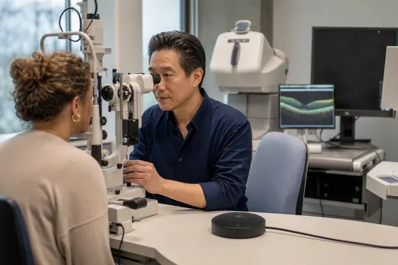
A structured report in your professional terminology
From care encounter to report
Hysio turns spoken information into a report. Choose the report format for the conversation you are documenting.
Document
Record the conversation and clearly state your own findings and agreed actions.
AI assistants
You stay in control of what gets saved.
In your workday
More attention for the conversation, less typing afterwards.
State relevant information, your own findings and agreed actions aloud so you can find them in your report.
The information appears under the headings of your chosen report format, so you can review each section.
Check names, facts and agreed actions. Edit the text where needed before using the report in your records.
Try it yourself
Try Hysio free for 14 days or schedule a personal demo.
For your team
Quick answer
For vision and eye health, with a dedicated place per eye for visual acuity, intraocular pressure, slit lamp, fundus and procedures.
This is how Hysio works in an ophthalmology consultation, examination or check-up. The answers are based on the documentation formats for the ophthalmologist and the sources of the specialty.
When it comes to the request for help, fundus findings usually belong in the Ophthalmology Consultation or the Ophthalmology Intake, the formats for a first contact. There, fundus findings get a place alongside visual acuity and intraocular pressure, with the source that supplied them. If the finding comes from targeted follow-up examination, the Ophthalmology Examination Report is a better fit; if a procedure follows, the Procedure Report is the right fit. Slit-lamp examination and perimetry get their own place in the same structure, so the ophthalmologist can see at once which examination supports which finding. [2] [3] [4] [5] [6] [1] [12]
Intraocular pressure and slit-lamp examination each get their own heading in the report, with the source and time of measurement. Intraocular pressure usually belongs with the objective findings, slit-lamp examination with the physical examination. Set out visual acuity and fundus findings in the same order, so a colleague can follow the course of the examination without assumptions. You add perimetry once it has been performed, stating the instrument and the date, so the origin of each piece of data stays recognisable. [2] [1] [11]
You document perimetry as a separate part of the examination report, with the instrument, date and the supplied result. Link the outcome to the reason the test was performed, so the record path from request for help to finding stays recognisable. You set out visual acuity and intraocular pressure alongside it in the same structure, without rephrasing the perimetry result into a conclusion. Slit-lamp examination and fundus findings get their own line, so that you as the ophthalmologist assess the result yourself before it is finalised in the record. [11] [1] [2]
You only include slit-lamp examination as the source supplies it, without extra details that are not needed for this encounter. Record who gave the finding and what it is used for, so the privacy choice fits this specific encounter. The same applies to visual acuity, intraocular pressure, fundus findings and perimetry: only what was measured or said and what is needed for the record. You record consent for sharing separately, apart from the clinical finding itself. [9] [16] [1]
The price of Hysio does not depend on visual acuity or any other specific part of the examination, but on the plan you choose. The current amounts for Starter, Pro and Unlimited are on the central pricing page, including the annual discount. With voice-driven reporting, intraocular pressure, slit-lamp examination, fundus findings and perimetry all fall under the same source-bound way of working, with no separate costs per part of the examination. A free trial lets you assess in advance whether the structure fits your practice before you choose a plan. [7] [8]
Your own template for fundus findings can be a better fit when your practice already uses a fixed layout that does not correspond one-to-one with the Hysio structure. In that case, compare which parts, such as visual acuity, intraocular pressure and slit-lamp examination, appear in both layouts and where differences arise. Perimetry stays source-bound in every layout: the result itself does not change, only the place where it is noted. Choose the route that leads your team to a verifiable report the fastest. [1] [11] [12]
Place intraocular pressure next to the original source, for example the dictation or the measurement, and compare whether the value, unit and moment match. A fixed format makes the text clear, but says nothing about the accuracy of the measurement itself. Make the same comparison for slit-lamp examination and fundus findings: is the finding literally in the source, or has it been summarised? You check visual acuity and perimetry in the same way before you consider the report final. [1] [12] [10]
Discuss with the patient why perimetry is being performed and note what the patient asks or understands about it. In the conversation with the patient, keep the explanation, your own observation and the professional conclusion separate. The ophthalmologist determines what the finding means personally for this patient and checks the text before saving. [1] [2] [11]
Hysio organises slit-lamp examination in a fixed structure that follows the source, instead of free text that you have to lay out again each time. The format links the finding to the moment and context of the encounter, so visual acuity, intraocular pressure and fundus findings stay recognisable alongside it. Perimetry gets the same treatment: Hysio puts the result in the right place, but does not assess its content. You remain the one who interprets the findings and decides on the follow-up. [1] [2] [11]
Assessment of the report structure centres on visual acuity in one straightforward encounter, for example a single Ophthalmology Consultation, before the structure is assessed more widely. At the input step, also note intraocular pressure and slit-lamp examination, so you see in one go how the structure sets several parts of the examination side by side. Agree within your team who reads through the first reports. You add fundus findings and perimetry once they have been performed, so you can assess the build-up of the report step by step. [8] [9] [1]
Hysio Ophthalmology Consultation fits a regular encounter in which you bring together the reason for the visit, the examination and the follow-up discussed in one report. The format sets visual acuity, intraocular pressure and slit-lamp examination side by side, with the source that supplies each piece of data. Fundus findings and perimetry get their own place once they have been performed during the consultation. If the situation is more of a first introduction, the Ophthalmology Intake is the more obvious choice; if it concerns a separate examination result, the Examination Report is a better fit. [2] [1] [12]
Hysio Ophthalmology Intake fits the first contact, in which the request for help, history and context of the complaint are recorded. The format sets visual acuity and intraocular pressure side by side with the source that supplied them, and leaves room for slit-lamp examination once it has been done during the intake. Fundus findings and perimetry usually only follow at a follow-up encounter. If there is already a record with earlier findings, the Ophthalmology Consultation or the Examination Report fits that follow-up better. [3] [1] [12]
Hysio Ophthalmology Examination Report fits an encounter in which a specific examination, such as perimetry or slit-lamp examination, determines the reason and the conclusion. The format sets the result alongside visual acuity and intraocular pressure, with the source and time of measurement. Fundus findings get their own line once they belong to the examination. If it concerns a broader first consultation without a specific examination, the Ophthalmology Consultation or the Intake is a better fit for that moment. [4] [1] [12]
Hysio Ophthalmology Procedure Report fits an encounter in which an intervention or procedure was carried out, with the reason, execution, findings and aftercare as fixed parts. The format sets intraocular pressure and slit-lamp examination side by side as part of the preparation or check-up around the procedure. Fundus findings and perimetry get a place once they belong to the intervention. If it concerns a regular consultation without a procedure, the Ophthalmology Consultation is a better fit for that encounter. [5] [1] [12]
Hysio Ophthalmology SOAP Note fits an encounter that you want to document according to Subjective, Objective, Assessment and Plan, for example a results or check-up discussion. Visual acuity and intraocular pressure go under Objective, where slit-lamp examination and fundus findings get their own line. You add perimetry once it has been performed, with the result alongside the assessment you give as the ophthalmologist. If the encounter is more of a first introduction, the Ophthalmology Intake is the more obvious choice. [6] [1] [12]
Which format fits visual acuity best depends on the moment in the encounter. If it is a first measurement at registration, visual acuity belongs in the Intake or the Consultation; if it is part of a targeted examination, the Examination Report is a better fit. If visual acuity follows a procedure, the Procedure Report is the right fit; if you discuss a result according to Subjective, Objective, Assessment and Plan, the SOAP Note is the right fit. Intraocular pressure, slit-lamp examination and fundus findings follow the same logic of choice for each moment in the encounter. [1] [2] [12]
You choose an intake at the first contact, in which the request for help and the patient's history are recorded. Note the laterality, instrument, time of measurement and findings with their source. The ophthalmologist assesses the outcome and determines the follow-up; Hysio does not add any interpretation. [1] [2] [12]
Once the results are in, intraocular pressure should be recorded with the measured value, the instrument, the date and the source that supplied the measurement. Also briefly describe the reason for the measurement and the follow-up discussed, without adding a judgement about the cause. You note slit-lamp examination and fundus findings in the same way: factually and with their origin. You add perimetry when it belongs to the same result, so the ophthalmologist finds the complete picture in one report. [1] [2] [12]
In a handover, perimetry should include the result itself, the instrument, the date and who performed or assessed the test. Also state the reason for the handover and to whom it is addressed, so the recipient understands the context without having to read through the full history. You share intraocular pressure, slit-lamp examination and fundus findings in the same way: briefly, traceably and with the source. Before sending, check whether consent for sharing this information has been recorded. [1] [2] [12]
For slit-lamp examination, keep what the patient tells you, what you observe yourself and what the ophthalmologist interprets professionally separate. Note the laterality, the method used and the moment of examination. The ophthalmologist checks the source and determines which conclusion and next step go into the record. [1] [2] [12]
Record in the patient's own words what the patient tells you, which information comes from the patient and what comes from the examination source. Note the source, moment and laterality separately. The ophthalmologist assesses the findings and determines the follow-up without presenting patient information as their own observation. [1] [12]
For each ophthalmic value, note whether it comes from the patient, from an instrument or from another source. Record the instrument, time of measurement and laterality. The ophthalmologist assesses how it fits together, and Hysio does not turn the measured value into an independent diagnosis or treatment choice. [1] [16] [12]
For fundus findings, note the instrument, the date and the time of the measurement, together with the laterality. Place the finding next to earlier measurements when they are available, so a course becomes visible without you already attaching a conclusion to it. Visual acuity, intraocular pressure, slit-lamp examination and perimetry follow the same approach. You determine the professional meaning of the measurement yourself; Hysio only sets out the data clearly. [1] [12]
Link intraocular pressure to the result by connecting the measured value directly to the examination in which it was taken, including the date and source. With voice-driven reporting, you state your own assessment in a separate sentence, so measurement and interpretation stay apart. You link slit-lamp examination and fundus findings to their result in the same way. You add perimetry once it belongs to the same episode, so the complete examination picture comes together in one report. [1] [12] [16]
No, Hysio does not assess images. For perimetry, Hysio uses a supplied report or a result you have dictated, but does not interpret scans, photos or test results itself. Intraocular pressure, slit-lamp examination and fundus findings are likewise only included as the source supplies them. You remain the one who assesses imaging and determines what it means for visual acuity and the follow-up; Hysio only puts that assessment into the report, tied to the source. [11] [10] [12]
If you want to record an explanation in plain language for a conversation with the patient, note what the patient asked, which explanation you gave and which agreement followed from it. Keep the source of the explanation and your professional conclusion separate. The ophthalmologist checks the text before it is shared according to local agreements. [1] [12]
Note whether the information comes from the patient and which information a relative or healthcare professional contributed. Also record when the patient is not able to indicate something clearly. The ophthalmologist assesses the meaning and keeps the source, observation and professional conclusion clearly separate. [1] [16] [12]
For a multidisciplinary meeting, the team mainly needs the result of the fundus findings, the time of measurement and your initial view of the follow-up, in language that someone who is not an ophthalmologist can also follow. State which discipline supplied the finding and who takes up the follow-up. You describe visual acuity, intraocular pressure, slit-lamp examination and perimetry in the same concise way. This lets the team quickly judge whether a finding has consequences for their own part of the care, without searching the full record. [1] [2] [16]
A useful example case for intraocular pressure describes a real encounter: the reason, the measured value, and the follow-up that you as the ophthalmologist discussed. Show how intraocular pressure ends up in the report alongside visual acuity and slit-lamp examination, without inventing a diagnosis or cause that does not come from the source. Fundus findings and perimetry can play a role in the same case when they belong to the same encounter. An example therefore shows the structure, not a fixed medical outcome. [1] [12] [2]
Limit a handover about perimetry to the result, the instrument, the date and the purpose of the handover, without additional interpretation that does not come from the source. State to whom the handover is addressed and let that person trace visual acuity and any other findings from the record themselves. You share intraocular pressure, slit-lamp examination and fundus findings in the same source-bound way. Before sending, check whether consent for this handover has been recorded. [11] [9] [16] [12]
No, Hysio does not make a diagnosis for perimetry. The format sets out the result, the instrument and the date clearly, but does not assess what the outcome means for the ophthalmic conclusion. Intraocular pressure, slit-lamp examination and fundus findings get the same treatment: Hysio organises the finding, but does not interpret it. Visual acuity and the eventual follow-up stay entirely with you as the ophthalmologist, who weighs the findings together and records the conclusion. [10] [1] [12]
You check slit-lamp examination before the report is saved; Hysio structures the text, but does not take over that professional check. Read the finding back next to the source and correct anything that is not right or is incomplete before you make the report final. Visual acuity, intraocular pressure, fundus findings and perimetry go through the same check. That way, final responsibility for the content of the record stays with you, even when the structure has been drawn up automatically. [1] [10] [16]
If visual acuity is missing from the encounter, you mark it in the report as missing or not discussed, instead of adding a value or estimate that does not come from the source. The same rule applies to intraocular pressure, slit-lamp examination, fundus findings and perimetry: an empty data field stays empty. That way, a colleague who reads the record later can clearly see what was actually measured during this encounter and what still needs to be done. [1] [10] [12]
No, Hysio is not intended for the acute assessment of fundus findings. In an acute situation, you first decide yourself on the urgency and the follow-up; Hysio only records afterwards what happened and which findings were made. Visual acuity, intraocular pressure, slit-lamp examination and perimetry follow the same order: first the acute assessment, then the source-bound documentation. So do not use Hysio as an aid to determine for yourself what is going on during an acute assessment. [10] [9] [12]
Record the consent the patient gave for this, with the purpose, the moment and the recipient. In addition, note which result or finding actually comes from the source. The ophthalmologist assesses the content and determines which passage is saved or shared according to local agreements. [9] [16] [1]
No, Hysio does not make a treatment plan around perimetry. The format puts the result under a clear heading, but does not choose a next step, medication or intervention. Intraocular pressure, slit-lamp examination and fundus findings have the same limit: Hysio organises the finding, you determine the plan. You note visual acuity and the follow-up discussed yourself in your own words, so the plan demonstrably remains your professional choice and not automatically generated advice. [10] [11] [12]
Leave doubt about slit-lamp examination visible in the report, for example by noting that a finding was not clear-cut or calls for further examination, instead of making the text more definite than the source allows. The same approach applies to visual acuity, intraocular pressure, fundus findings and perimetry: uncertainty belongs in the report, not only in your head. That way, a colleague who reads the record later can recognise the open question and come back to it themselves. [1] [12] [10]
When sources on visual acuity differ, for example an older measurement versus your own examination today, you keep both readings separately with their own origin and date. Do not automatically choose one version as correct; note the difference and explain what you established yourself. Intraocular pressure, slit-lamp examination, fundus findings and perimetry follow the same approach. That way, it stays visible why a value in the record has changed, instead of an older measurement silently disappearing. [1] [16] [12]
Record medication and fundus findings exactly as the source supplies them, including the dosage, unit and the moment of measurement or prescription. Put both under their own heading, so medication is not accidentally read as part of the fundus finding itself. Visual acuity, intraocular pressure, slit-lamp examination and perimetry follow the same separation between data. You assess yourself whether medication and finding are related; Hysio only sets them out clearly side by side. [1] [12] [10]
Before saving, read intraocular pressure back next to the original source and the date, and check whether the value and unit are correct. Correct anything that does not match before you make the report final; the report enters your record only after you have checked and saved it. Slit-lamp examination, fundus findings, visual acuity and perimetry go through the same final check. Admin with AI takes over the writing out, but the last step before saving remains your own deliberate assessment. [1] [9] [10] [16]
Record what you discussed with the patient, which ophthalmic finding came from the source and which agreement was made. Note the laterality and the moment of the encounter. The ophthalmologist checks the text and determines which conclusion or next step goes to the record. [11] [9] [16] [2]
For perimetry, you mainly need an agreement within the team about fixed headings and shared terms, so everyone finds the same result in the same way. Also record who may enter the result and who reads through or approves it afterwards. Intraocular pressure, slit-lamp examination, fundus findings and visual acuity call for the same agreements. Without such a shared way of working, differences arise between colleagues that make reading the record unnecessarily harder. [1] [2] [11]
Start with slit-lamp examination in a clearly defined encounter, for example a single consultation, and then gather for yourself what does not yet work in the set-up. Only extend to visual acuity, intraocular pressure, fundus findings and perimetry once that first trial shows that the structure fits your way of working. A small trial shows whether the layout matches your own use of language, without you having to overhaul your whole practice straight away. [8] [9] [1]
Adapt a report on visual acuity by aligning the headings and order with the agreements that already apply in your practice, for example the layout your colleagues also use. You check intraocular pressure against the same local authorisations: who may record or change which value. Slit-lamp examination, fundus findings and perimetry follow the same adaptation. Have a designated colleague read through the first adapted reports, so the local agreements have been properly incorporated before you work more widely. [11] [1] [12]
For each eye, state what you saw on fundoscopy: disc, macula, vessels and periphery. Hysio puts it with the correct eye and keeps visual acuity, intraocular pressure and slit-lamp examination each in their own place. You mention perimetry or an OCT with the date of the examination. You check that left and right are correct before you save. [1] [12] [2]
Measure time savings around intraocular pressure by comparing within your team how long documentation took before and after using a fixed structure, including the time for checking and corrections. Make the same measurement for slit-lamp examination, fundus findings, visual acuity and perimetry. A reliable measurement looks at your own practice, not at a generally promised percentage. Always give the quality of the report priority over faster but less careful recording. [1] [11] [8]
There are no separate trial conditions for perimetry; Hysio's central free trial applies to all parts of the examination at once, including intraocular pressure, slit-lamp examination, fundus findings and visual acuity. During the free trial, you can assess for yourself whether the structure for perimetry results matches how you normally draw up reports. The current length and conditions of the free trial are on the central page about it, together with the prices after the trial ends. [8] [7] [9]
The pricing page describes what a Hysio plan costs and which conditions apply. Slit-lamp examination, visual acuity measurements and other ophthalmic data have no separate price within that plan. The legal pages explain data processing and responsibility. Use that explanation when choosing the platform, while the content and assessment of the ophthalmic record remain your professional responsibility. [7] [9] [10]
Record when different ophthalmologists treat the same patient, which source belongs to which encounter and which agreement was made. Keep your own observation and the professional conclusion separate. The treating ophthalmologist checks the handover and determines what the next colleague needs. [1] [11] [12]
Break a complex case around fundus findings down by encounter and source: which finding belongs to which consultation, and who the information came from. Then organise intraocular pressure, slit-lamp examination, visual acuity and perimetry in the same order, without adding an extra conclusion that goes beyond the individual findings. That way, a case with several encounters and parts of the examination stays readable, and you as the ophthalmologist decide yourself how the separate findings together form a picture. [1] [12] [2] [10]
Hysio creates the report. You remain responsible for what you save in the record.