Document
Keep your attention on the care encounter.
Record the conversation and clearly state your own findings and agreed actions.
Hysio Radiology
You dictate clinical question, findings and conclusion. Hysio puts them into a radiology report, with the comparison with earlier imaging in its own paragraph.
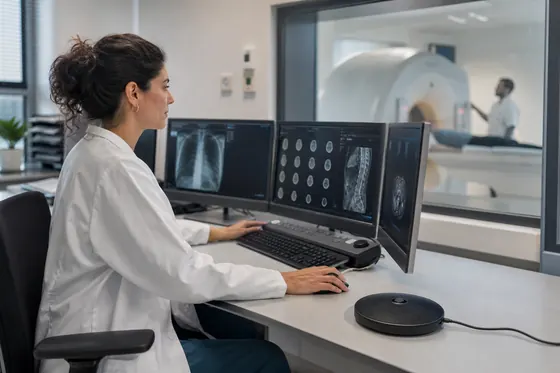
A structured report in your professional terminology
From care encounter to report
Hysio turns spoken information into a report. Choose the report format for the conversation you are documenting.
Document
Record the conversation and clearly state your own findings and agreed actions.
AI assistants
You stay in control of what gets saved.
In your workday
More attention for the conversation, less typing afterwards.
State relevant information, your own findings and agreed actions aloud so you can find them in your report.
The information appears under the headings of your chosen report format, so you can review each section.
Check names, facts and agreed actions. Edit the text where needed before using the report in your records.
Try it yourself
Try Hysio free for 14 days or schedule a personal demo.
For your team
Quick answer
For imaging and image-guided procedures, with a dedicated place for clinical question, technique, findings and comparison.
Read what Hysio does for a radiology report or an interventional radiology procedure. For each documentation format you can see what goes into it and where the line is.
For the request for help, the documentation format that fits is the one that matches the comparison material and the chosen imaging modality. Hysio links the clinical question and modality to the contact, while the assessment of the imaging stays with you. You place comparison and clinical information side by side and write down only what the source states for this contact. This keeps it traceable which prior examination was used for the comparison. [2] [3] [1] [11]
You organise imaging modality and findings under fixed headings in the record, with clinical information and the clinical question right next to them. Each item keeps its origin, so you can later quickly find where a value came from. You describe comparison in the words of the source, not in an interpretation of your own. This keeps the administrative structure clear and source-bound. Image assessment by the radiologist precedes diagnostic dictation. Check the organised report text before saving it. [2] [1] [8]
You record clinical information along the record pathway that belongs to your route in the medical record system, alongside the clinical question, comparison and the source. Hysio works from that clinical information and only connects it to a follow-up after you have assessed the contact. Check the clinical question and the source before you save the text. This way the record pathway fits your existing way of working, without Hysio choosing a route itself. [8] [1] [2]
When making the privacy choice, you place the clinical question and imaging modality side by side and keep the imaging with a clear origin. The privacy choice gets its own source alongside the comparison material, and you record how clinical information was discussed. Limit the note to what this one contact needs, and record consent for sharing clinical information separately. This way only what actually proves relevant for this contact remains visible. [6] [9] [1]
The price for Hysio is shown on the central pricing page and applies to every radiologist, whether you work with the clinical question, imaging modality or comparison. On that page you will find the current rates per plan and the conditions for a free trial. The price depends on the plan you choose, not on the imaging modality. [4] [5]
A template of your own for comparison can be a better fit when your team already uses a fixed layout that does not correspond one to one with the Hysio format. In that case, link the clinical question to imaging modality and the contact, and keep clinical information traceable to the source. Compare your own template with Hysio before you choose, and write only what the source states for that contact. This way you decide yourself when another route is a better fit. [1] [8] [11]
During the source check, you compare the report with the dictation and check imaging modality and comparison word for word against the source. Record clinical information and the clinical question with their origin, so that discrepancies stand out. A fixed format does not automatically make the assessment of the imaging correct; that check remains yours. This keeps the source check an explicit step rather than an assumption. [1] [11] [7]
If there is patient contact, you focus on the patient: you write up clinical information from what was said during that contact, not from an assumption. You place imaging modality and comparison alongside it, with the source included. Check the clinical question before you finalise the note, so that the assessment of the imaging matches what the patient has indicated. This keeps the patient's perspective recognisable in the record. [1] [2] [8]
Hysio organises the clinical question, imaging modality and comparison clearly and links each item to the source it comes from. You remain the one who assesses clinical information and decides what is useful for the final report text. So the difference is not in the conclusion, but in how the report text is organised. [1] [2] [8]
At the input step, begin with one recognisable contact in which the clinical question and clinical information are clearly recorded. Record comparison and imaging modality with their available origin. Before diagnostic dictation takes place, the radiologist assesses the images. Review the organised report text before you save it. Agree with your team who reviews the outcome before you use the approach more widely. This way use grows step by step from one verified example. [5] [6] [1]
Hysio Radiology Report suits the contact for a routine radiological examination: the format brings together imaging modality, comparison and the follow-up of that contact. You record clinical information and the clinical question alongside it, with the source included, and you assess the imaging yourself. The separate procedure format is intended for an interventional radiology procedure. With voice-driven reporting you link the comparison to the clinical question; saving waits for your check. [2] [1] [11]
Hysio Interventional Radiology Procedure Report suits the interventional radiology procedure itself, with the clinical question and imaging modality recorded alongside the intervention performed. The format keeps clinical information with its origin, so that the reason for the procedure also remains traceable. The Radiology Report is intended for a routine report without an intervention. [3] [1] [11]
The format that fits depends on the contact: Hysio Radiology Report is intended for a routine examination, the procedure format for an intervention. Record clinical information and the clinical question with their available origin. The radiologist first assesses the images, before diagnostic dictation. Before saving, check that the organised report text is correct. Connect imaging modality and comparison to the source that this contact contains. This way the chosen format matches what was actually performed. [1] [2] [11]
At a first assessment, clinical information is recorded for the first time, with the clinical question and imaging modality as the starting point. In a follow-up contact you mainly describe what has changed since that first assessment, as with comparison. Link imaging modality to comparison and the contact, and write down only what the source states for that moment. This keeps the distinction between a first assessment and a follow-up recognisable. [1] [2] [11]
After the handover question, the reason, the procedure performed and the findings belong together, with the clinical question and imaging modality as their origin. Do not add details that do not belong with comparison or clinical information. The images are assessed by the radiologist before diagnostic dictation. You check the organised report text before saving it. This keeps the handover limited to what the contact actually produced. [1] [2] [11]
A handover includes the clinical question with the source it comes from, together with imaging modality and comparison. With voice-driven reporting too, you make the source, recipient and agreement visible, and you check consent before you share anything. You include clinical information only when the follow-up contact requires it. This keeps the handover limited and traceable. [1] [2] [11]
Keep the patient's account, observation and professional assessment apart when you record comparison. For each item, note the source role: from the patient, from a prior examination, or from your own assessment of the imaging. You place clinical information and the clinical question alongside it, with imaging modality as context. This keeps it clear which part of the report rests on which source. [1] [2] [11]
Compare imaging modality per contact on the same key points, so that a change is actually a change and not merely a difference in writing style. Name only changes that the source on the image interpretation actually supports. You include clinical information and the clinical question with their available origin. Connect comparison and the source to the moment of the result at which you make that assessment. [1] [2] [11]
At the results contact, record which result about clinical information was discussed, with the date and source included. Keep the explanation and the follow-up separate from the image interpretation itself, so that facts and interpretation do not get mixed up. You record clinical information and the clinical question side by side, with only what the source states for this contact. You link imaging modality to that clinical information. [1] [2] [11]
The length of the report follows from the contact and its purpose, not from a fixed rule. For comparison you describe only what has changed; for a first examination you write up clinical information and the clinical question in more detail. Imaging modality in the words of the source remains the guide. Diagnostic dictation follows the radiologist’s assessment of the images. Review the organised report text for accuracy before saving. [1] [2] [11]
In the history you record the request for help and the course in the words of the source, with the clinical question and imaging modality as their origin. You add comparison only if it was discussed during the contact, not as an addition of your own. You place clinical information alongside it, with the source clearly stated. The radiologist’s assessment of the images comes before diagnostic dictation. Go through the organised report text before saving. [1] [11]
Record who provided the clinical question: the patient, a relative or a referrer, and separate that from your own observation. This keeps imaging modality traceable to the right source within the image interpretation. You place comparison and clinical information alongside it with only what that source states. Before diagnostic dictation, the radiologist assesses the images. Check the organised text of the report before saving it. [1] [9] [11]
For comparison, record the instrument, the unit and the time of measurement, so that the value remains traceable. You record the clinical question and imaging modality with their origin, alongside clinical information in the words of the source. You determine the professional meaning of that measurement within the image interpretation, not Hysio. This way the measured value stays a fact and the interpretation stays your assessment. [1] [11]
Link imaging modality to the examination, the date and the source, so that the result remains traceable. You place clinical information and the clinical question alongside that examination result. Give your own assessment of comparison its own place in the report, separate from the image interpretation provided. This keeps fact and assessment separate. [1] [11] [9]
No, Hysio does not assess images itself. Hysio does incorporate a report on the imaging that has been provided, with imaging modality and comparison alongside it and the source clearly stated. You record clinical information and the clinical question as they were discussed. The source remains decisive for the image interpretation; you make that assessment yourself. [8] [7] [11]
Use the professional terms that are common within your team, and define abbreviations once so that comparison and clinical information remain unambiguous to read. You describe the clinical question and imaging modality in the words of the source. Check whether colleagues read the image interpretation the same way you do. This prevents confusion between disciplines. [1] [11]
Always state who the clinical question comes from: the patient, a relative or a referrer. Record consent for sharing imaging modality separately, before you share anything. You place comparison and clinical information alongside it, with only what that source states. This keeps both origin and consent traceable in the contact. [1] [9] [11]
For a team meeting, you make visible which discipline provided the comparison and who takes on the follow-up. You record the clinical question and imaging modality with their origin, alongside clinical information in the words of the source. Keep that clinical information understandable for colleagues outside radiology, without rewriting the image interpretation itself. This keeps the team meeting focused on what each discipline needs. [1] [2] [9]
An example shows the real contact: which imaging modality was used, what the comparison yielded and what the follow-up was. You place clinical information and the clinical question alongside that example case, with the source included. Do not invent a diagnosis that was not recorded in the underlying contact. This keeps an example recognisable as an illustration, not as a new fact about the image interpretation. [1] [11] [2]
Limit a handover about clinical information to source and purpose, so that the recipient can trace the clinical question themselves. You place imaging modality and comparison alongside it, with the origin included. Record how that clinical information was discussed, without adding an interpretation of your own to the image interpretation. This keeps the handover factual and compact. [8] [6] [9] [11]
No, Hysio does not make a diagnosis and does not set an indication. Hysio organises clinical information in the words of the source, alongside the clinical question, imaging modality and comparison with their origin included. The radiologist determines the meaning of that information within the image interpretation. So always assess the imaging yourself, however clearly the record has been organised. [7] [1] [11]
You review the text for the clinical question, findings and the follow-up stated, and correct whatever does not match the source. Record the modality used and the time of measurement only when they are in the source. The radiologist determines the professional assessment and the follow-up; Hysio does not add a diagnosis or interpretation. [1] [7] [9]
If the clinical question is missing, mark that explicitly instead of making something up yourself. You place comparison and clinical information alongside it with the source that does state something about it. Do not add a date, value or conclusion that was not discussed in the contact. This keeps it clear what is really missing within the image interpretation, instead of it going unnoticed. [1] [7] [11]
No, do not use Hysio for triage or during the acute assessment itself. Record the clinical question and imaging modality only after that acute assessment, with their available origin. You record clinical information in a source-bound way as soon as the situation allows, alongside comparison. Always assess the imaging yourself; Hysio only organises afterwards what proves relevant for the image interpretation. [7] [6] [11]
Record which information was shared and for what reason, so that afterwards it remains traceable what the recipient received. Include the source, the moment and the agreed follow-up action. The radiologist checks the text before handover and decides which information may be provided under local agreements. [6] [9] [1]
No, Hysio does not choose a treatment or an operation. Hysio organises clinical information under a heading, with the clinical question in the words of the source alongside it. You record imaging modality and comparison with their origin included, so that the context remains visible. The radiologist oversees the image interpretation and the follow-up pathway. [7] [8] [11]
Leave doubt about the comparison recognisable instead of smoothing it over. Do not make clinical information more definite than the source allows, and record the open question separately. You place the clinical question and imaging modality alongside it, with the source included. That way voice-driven admin does not hide doubt behind wording that is too definite. [1] [11] [7]
Keep both readings when sources on the clinical question differ, and record their origin separately. You place comparison and clinical information alongside them, with imaging modality as context. Only then assess which reading carries the most weight for the image interpretation. This way neither source disappears from the record. [1] [9] [11]
For each value, state the time of measurement and the source that provided it, so that a later report does not silently replace the same value. Also record which clinical question and imaging modality belong with the value. The radiologist assesses the finding and the follow-up; Hysio draws no independent conclusion from it. [1] [11] [7]
At the final check, read the text on imaging modality back alongside the source and date, before you save it. You place clinical information and the clinical question side by side, with only what the source states. Correct discrepancies in comparison before the image interpretation is recorded definitively. This makes the final check the last step, not a formality. [1] [6] [7] [9]
For the team handover, agree who reviews the text on imaging modality. You place clinical information and the clinical question side by side, with a source alongside comparison. Decide in advance which information the follow-up contact needs, and set the rest apart within the image interpretation. This keeps the team handover short and focused on what the next colleague needs. [8] [6] [9] [2]
Within your team, agree on fixed headings for clinical information and shared terms for the clinical question. Record imaging modality and comparison with their available origin, and record who ultimately approves the image interpretation. The radiologist assesses the images first; diagnostic dictation follows. Before saving, review the organised text of the report. This way everyone in the team works from the same agreements. [1] [2] [8]
Start with comparison in one clearly defined contact, so that you can quickly see whether the format is right. Link the clinical question to imaging modality and that one contact, and collect corrections to clinical information before you go further. Write only what the source states. Only expand voice-driven reporting once the description of your image interpretation in the report is right. [5] [6] [1]
Check the clinical question against the headings your team already uses, and imaging modality against the applicable authorisations. You describe comparison and clinical information with their origin included. Have the designated colleague check the image interpretation before you change anything. This way the report fits existing local agreements instead of replacing them. [8] [1] [11]
State the earlier examination with modality and date, and say what has changed since. Hysio puts the comparison in its own paragraph, next to clinical question and findings. If there is no earlier examination, you say so and the field stays empty. You authorise the report. [1] [11] [2]
Measure checking and corrections around imaging modality within your own team, not against a national average. You place clinical information and the clinical question side by side, with the source included. Always give the quality of the image interpretation priority over a promised time percentage. This keeps time savings a by-product, not the goal in itself. [1] [8] [5]
For clinical information, read the central trial conditions before you begin. You record imaging modality and comparison with their available origin, alongside the clinical question and the source. Use the free trial to assess whether the representation of the image interpretation in the report works in practice. Always assess the imaging yourself during that trial too. [5] [4] [6]
The Hysio pricing page provides information about subscription costs and the free trial. The legal pages describe how data is processed and the responsibilities that come with use. Radiological findings or an imaging modality are not separate pricing options. The platform terms help you assess the use of Hysio; you assess the clinical question, findings and conclusion in the radiology report yourself on the basis of the available sources. [4] [6] [7]
Within your team, agree how you write the clinical question, comparison and frequently used abbreviations. You record clinical information and imaging modality with their origin included. Keep those agreements understandable for every colleague who later reads the image interpretation. This prevents the same term from being used in two ways. [1] [8] [11]
In a complex case, split comparison into separate contacts and sources, instead of cramming everything into one note. Organise clinical information, findings and the follow-up without adding an extra conclusion to the image interpretation. You place the clinical question and imaging modality alongside it, with the source included. This keeps even a complex case clear. [1] [11] [2] [7]
Hysio creates the report. You remain responsible for what you save in the record.